
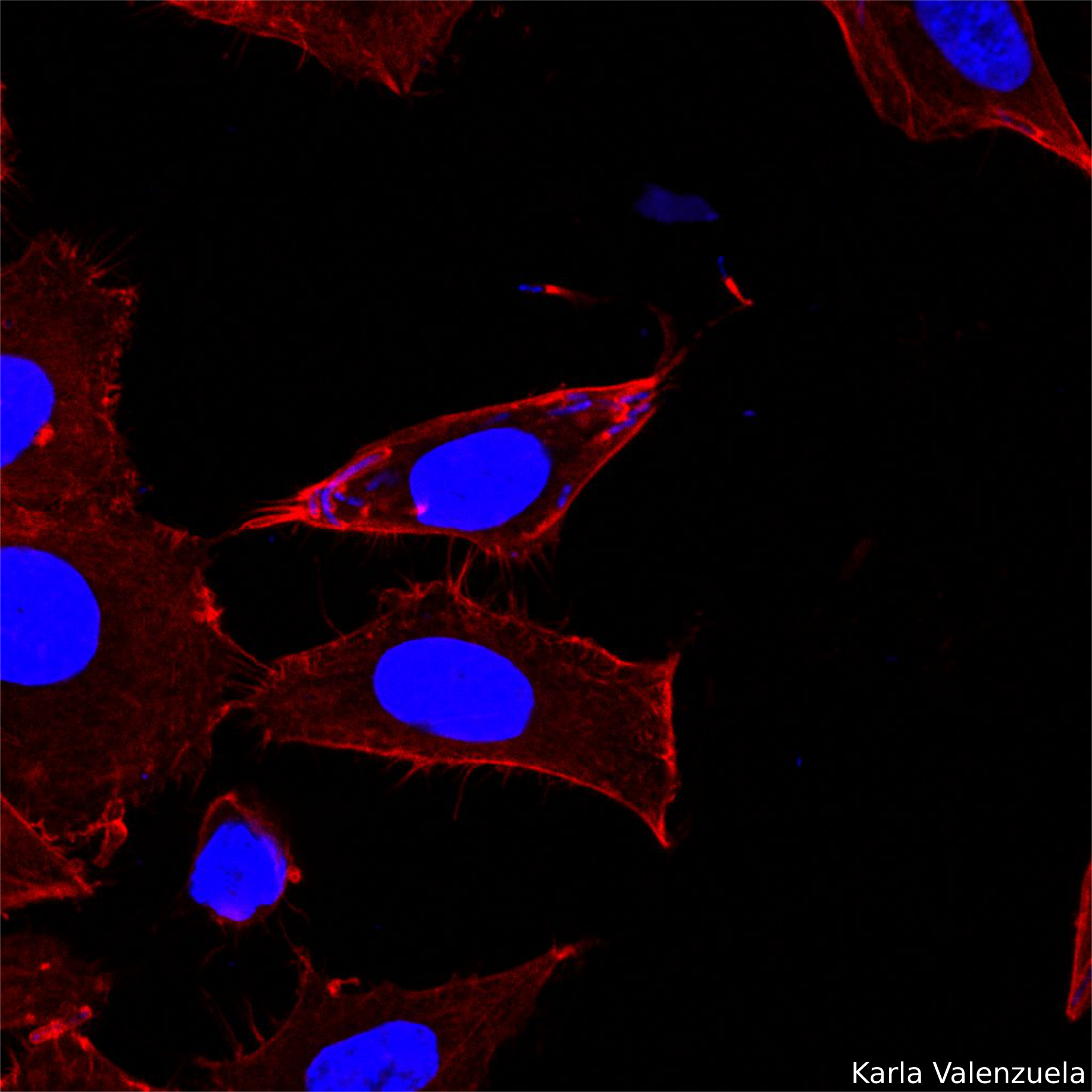
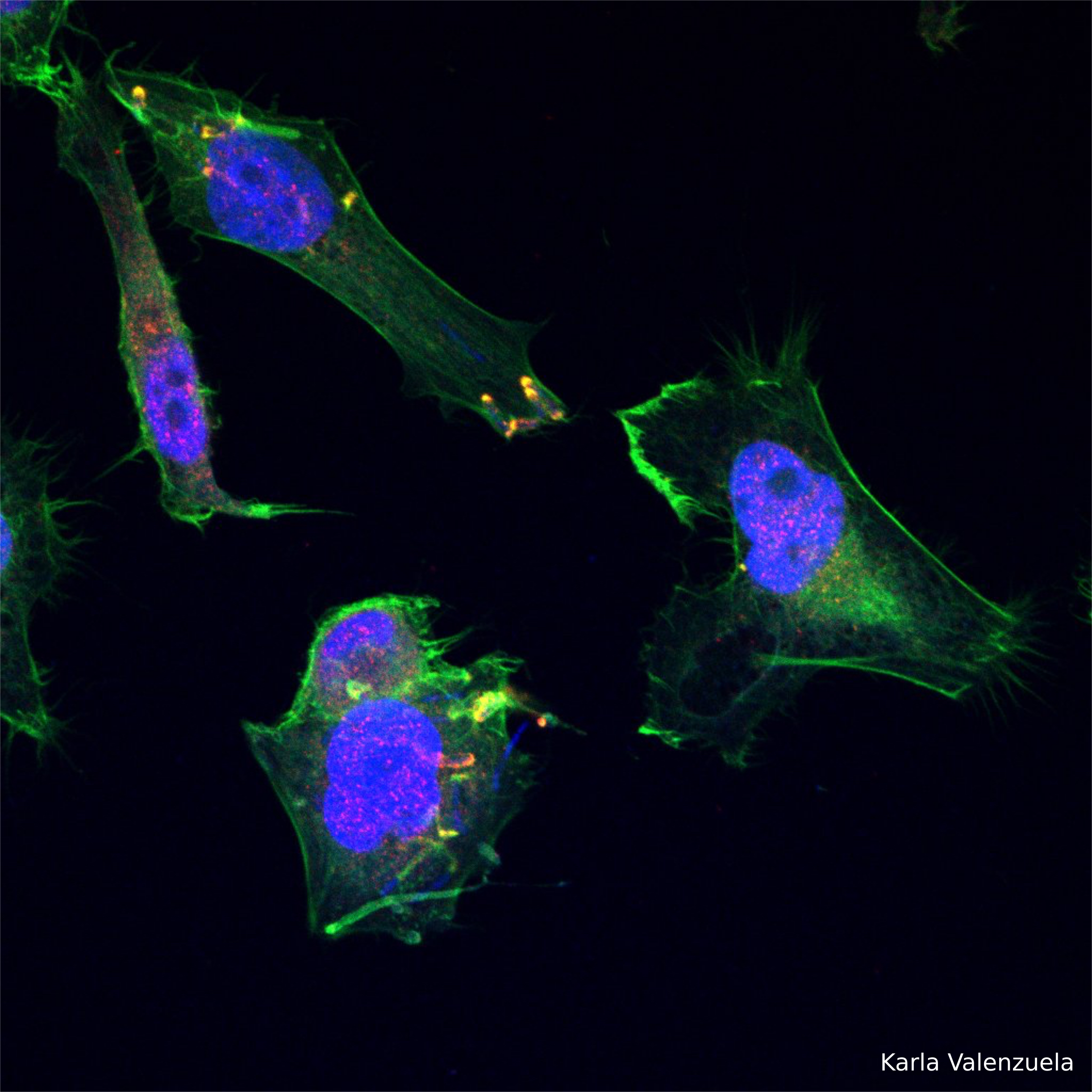
Actin tail assembly also enables Shigella to form a protrusion from the cell surface. The protrusion contacts the membrane of the adjacent cell allowing bacterial spreading. Red:Actin/Blue:DNA. Image taken with Zeiss LSM 710 Confocal Microscope.
Shigella activates the host Arp2/3 actin-nucleator complex triggering the formation of an actin tail on one pole of the bacterum. This tail serves to propel the bacteria within the host cell. Image shows co-localization of Arp3 to Shigella's actin tail. Red:Arp3/Green:Actin/Blue:DNA. Image taken with Zeiss LSM 710 Confocal Microscope.
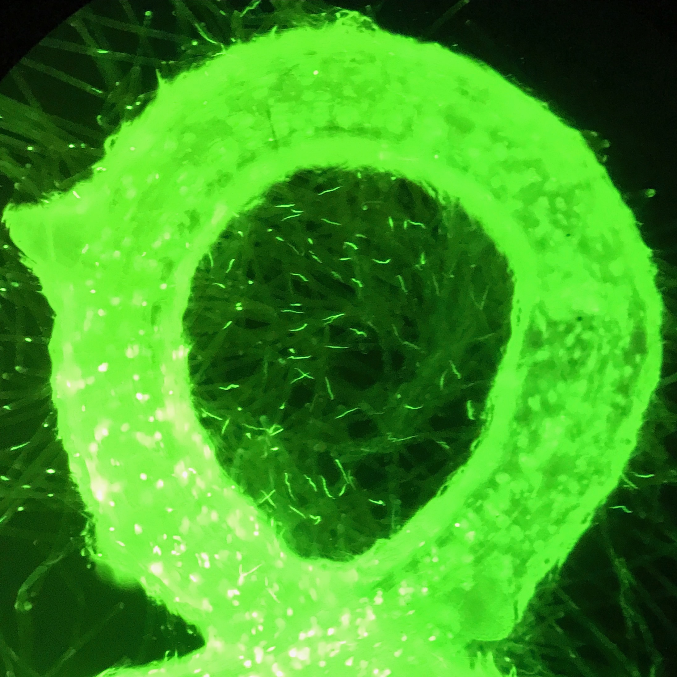
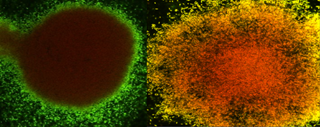
Confocal laser scanning microscopy (magnification power 63X) for biofilm grew under dynamicconditions, exposed to combination of alginate lyase and tobramycin. staining with Syto 61 red, emission fluorescence red from all cells, and Sytox green emission fluorescence green from dead cells, where the yellow fluorescent represented combination of both red and green channels. Control and treatment from left to right.


